Human Muscular System Diagram Photos and Premium High Res Pictures Biology Diagrams Learn about the three types of muscle tissue and the anatomy and function of skeletal muscles. Explore the 3D models of the muscular system and the naming of skeletal muscles. Learn about the muscular system, which is responsible for the movement of the human body. Explore detailed 3D anatomical illustrations of the skeletal, cardiovascular, digestive, endocrine, nervous, respiratory, immune, urinary, female reproductive, male reproductive, and integumentary systems. Explore the human muscular system like never before! This 3D visual journey covers muscle anatomy, functions, and movement in an easy-to-understand way. Perf
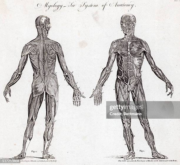
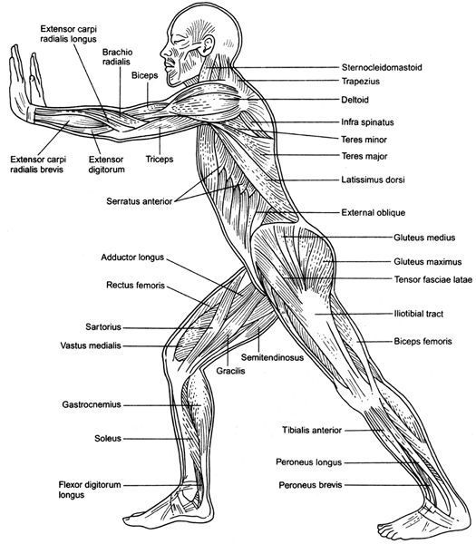
Muscular System Diagram. Here is the diagram of the human muscular system: Figure 5: Muscle chart showing the muscular system labeled where most muscles of the body are labeled in the form of a map of muscles. Image Credit: Wikimedia. Muscular System Physiology. Find out about the functions of the muscle tissues below.

Detailed 3D anatomical illustrations - Innerbody Biology Diagrams
Learn about the anatomy and physiology of muscles with Osmosis High-Yield Notes. See diagrams and illustrations of muscle fibers, sliding filament model, neuromuscular junction, and more.

Explore the muscular system in 3D with the BioDigital Human platform, a cloud-based virtual map of the human body. Learn about anatomy, disease and treatment with over 8,000 selectable structures and 850+ simulated conditions.
Muscle anatomy reference charts: Free PDF download Biology Diagrams
human muscle system, the muscles of the human body that work the skeletal system, that are under voluntary control, and that are concerned with movement, posture, and balance. Broadly considered, human muscle—like the muscles of all vertebrates—is often divided into striated muscle (or skeletal muscle), smooth muscle, and cardiac muscle.Smooth muscle is under involuntary control and is
Posted in Blog